Tag Archive for: liposomes

Publication on AWSensors technology

Acoustic detection of a mutation-specific Ligase Chain Reaction based on liposome amplification

Authors: Nikoletta Naoumi, Monica Araya-Farias, Maria Megariti, Lucile Alexandre, George Papadakis, Stephanie Descroix, and Electra Gizeli

Journal: Analyst

Abstract
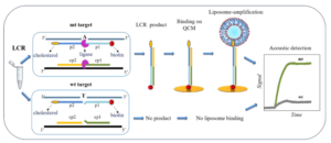
Single nucleotide variants (SNVs) play a crucial role in understanding genetic diseases, cancer development, and personalized medicine. However, existing ligase-based amplification and detection techniques, such as Rolling Circle Amplification and Ligase Detection Reaction, suffer from low efficiency and difficulties in product detection. To address these limitations, we propose a novel approach that combines Ligase Chain Reaction (LCR) with acoustic detection using highly dissipative liposomes. In our study, we are using LCR combined with biotin- and cholesterol-tagged primers to produce amplicons also modified at each end with a biotin and cholesterol molecule. We then apply the LCR mix without any purification directly on a neutravidin modified QCM device Au-surface, where the produced amplicons can bind specifically through the biotin end. To improve sensitivity, we finally introduce liposomes as signal enhancers. For demonstration, we used the detection of the BRAF V600E point mutation versus the wild-type allele, achieving an impressive detection limit of 220 aM of the mutant target in the presence of the same amount of the wild type. Finally, we combined the assay with a microfluidic fluidized bed DNA extraction technology, offering the potential for semi-automated detection of SNVs in patients’ crude samples. Overall, our LCR/acoustic method outperforms other LCR-based approaches and surface ligation biosensing techniques in terms of detection efficiency and time. It effectively overcomes challenges related to DNA detection, making it applicable in diverse fields, including genetic disease and pathogen detection.

You may read the full paper here.

Publication on AWSensors technology

C60-based Multivalent Glycoporphyrins Inhibit SARS-CoV-2 Specific Interaction with the DC-SIGN Transmembrane Receptor

Authors: Jennifer Patino-Alonso, Justo Cabrera-González, Javier Merino, Gema Nieto-Ortiz, Fátima Lasala, Jouma Katati, Carlos H. Bezerra da Cruz, Ajay K. Monnappa, Pablo Mateos-Gil, Ángeles Canales, Iván López-Montero, Beatriz M. Illescas, Rafael Delgado, and Nazario Martín

Journal: Small (2023)


Abstract

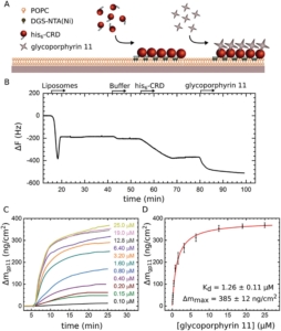
Since WHO has declared the COVID-19 outbreak a global pandemic, nearly seven million deaths have been reported. This efficient spread of Severe Acute Respiratory Syndrome Coronavirus-2 (SARS-CoV-2) is facilitated by the ability of the spike glycoprotein to bind multiple cell membrane receptors. Although ACE2 is identified as the main receptor for SARS-CoV-2, other receptors could play a role in viral entry. Among others, C-type lectins such as DC-SIGN are identified as efficient trans-receptor for SARS-CoV-2 infection, so the use of glycomimetics to inhibit the infection through the DC-SIGN blockade is an encouraging approach. In this regard, multivalent nanostructures based on glycosylated [60]fullerenes linked to a central porphyrin scaffold have been designed and tested against DC-SIGN-mediated SARS-CoV-2 infection. First results show an outstanding inhibition of the trans-infection up to 90%. In addition, a deeper understanding of nanostructure-receptor binding is achieved through microscopy techniques, high-resolution NMR experiments, Quartz Crystal Microbalance experiments, and molecular dynamic simulations.

You may read the full paper here.

Publication on AWSensors technology

Membrane insertion mechanism of the caveola coat protein Cavin1

Authors: Liu, K.-C., Pace, H., Larsson, E., Hossain, S., Kabedev, A., Shukla, A., Jerschabek, V., Mohan, J., Bergström, C. A. S., Bally, M., Schwieger, C., Hubert, M., & Lundmark, R.

Journal: PNAS (2022)


Abstract

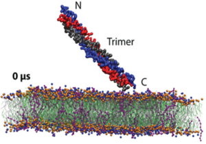
Caveolae are small plasma membrane invaginations, important for control of membrane tension, signaling cascades, and lipid sorting. The caveola coat protein Cavin1 is essential for shaping such high curvature membrane structures. Yet, a mechanistic understanding of how Cavin1 assembles at the membrane interface is lacking. Here, we used model membranes combined with biophysical dissection and computational modeling to show that Cavin1 inserts into membranes. We establish that initial phosphatidylinositol (4, 5) bisphosphate [PI(4,5)P2]–dependent membrane adsorption of the trimeric helical region 1 (HR1) of Cavin1 mediates the subsequent partial separation and membrane insertion of the individual helices. Insertion kinetics of HR1 is further enhanced by the presence of flanking negatively charged disordered regions, which was found important for the coassembly of Cavin1 with Caveolin1 in living cells. We propose that this intricate mechanism potentiates membrane curvature generation and facilitates dynamic rounds of assembly and disassembly of Cavin1 at the membrane.

You may read the full paper here.