Tag Archive for: SLB

Publication on AWSensors technology

V. cholerae MakA is a cholesterol-binding pore-forming toxin that induces non-canonical autophagy

Authors: Xiaotong Jia, Anastasia Knyazeva, Yu Zhang, Sergio Castro-Gonzalez, Shuhei Nakamura, Lars-Anders Carlson, Tamotsu Yoshimori, Dale P. Corkery, Yao-Wen Wu

JournalJ Cell Biol (2022)


Abstract

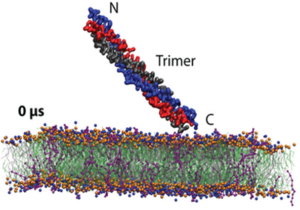
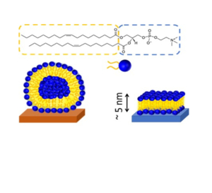
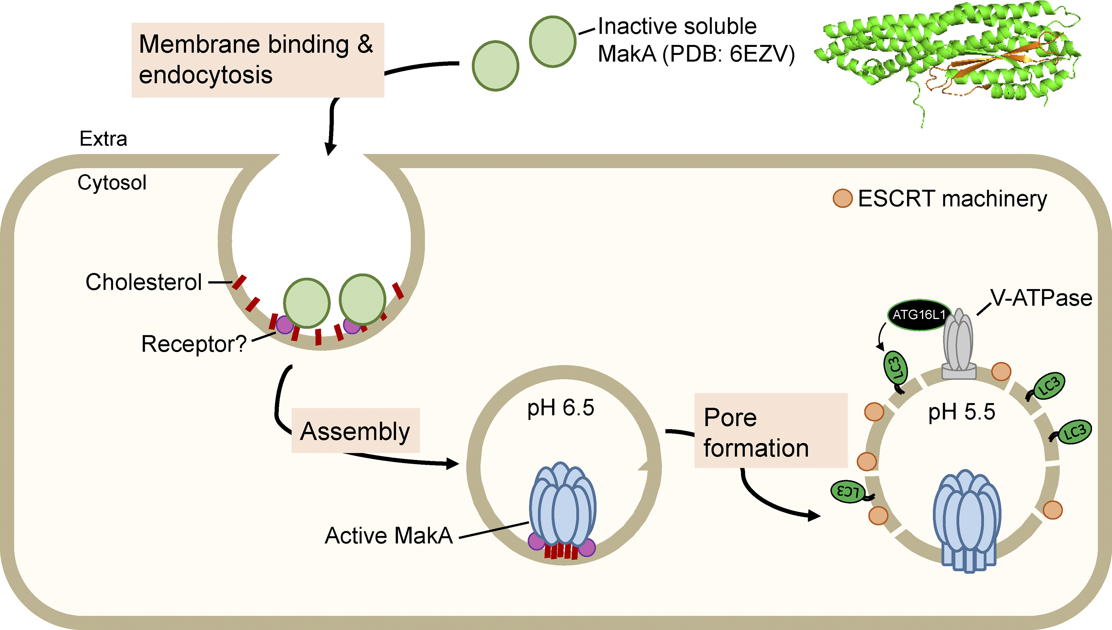
Pore-forming toxins (PFTs) are important virulence factors produced by many pathogenic bacteria. Here, we show that the Vibrio cholerae toxin MakA is a novel cholesterol-binding PFT that induces non-canonical autophagy in a pH-dependent manner. MakA specifically binds to cholesterol on the membrane at pH < 7. Cholesterol-binding leads to oligomerization of MakA on the membrane and pore formation at pH 5.5. Unlike other cholesterol-dependent cytolysins (CDCs) which bind cholesterol through a conserved cholesterol-binding motif (Thr-Leu pair), MakA contains an Ile-Ile pair that is essential for MakA-cholesterol interaction. Following internalization, endosomal acidification triggers MakA pore-assembly followed by ESCRT-mediated membrane repair and V-ATPase-dependent unconventional LC3 lipidation on the damaged endolysosomal membranes. These findings characterize a new cholesterol-binding toxin that forms pores in a pH-dependent manner and reveals the molecular mechanism of host autophagy manipulation.

You may read the full paper here.

Publication on AWSensors technology

Membrane insertion mechanism of the caveola coat protein Cavin1

Authors: Liu, K.-C., Pace, H., Larsson, E., Hossain, S., Kabedev, A., Shukla, A., Jerschabek, V., Mohan, J., Bergström, C. A. S., Bally, M., Schwieger, C., Hubert, M., & Lundmark, R.

Journal: PNAS (2022)


Abstract

Caveolae are small plasma membrane invaginations, important for control of membrane tension, signaling cascades, and lipid sorting. The caveola coat protein Cavin1 is essential for shaping such high curvature membrane structures. Yet, a mechanistic understanding of how Cavin1 assembles at the membrane interface is lacking. Here, we used model membranes combined with biophysical dissection and computational modeling to show that Cavin1 inserts into membranes. We establish that initial phosphatidylinositol (4, 5) bisphosphate [PI(4,5)P2]–dependent membrane adsorption of the trimeric helical region 1 (HR1) of Cavin1 mediates the subsequent partial separation and membrane insertion of the individual helices. Insertion kinetics of HR1 is further enhanced by the presence of flanking negatively charged disordered regions, which was found important for the coassembly of Cavin1 with Caveolin1 in living cells. We propose that this intricate mechanism potentiates membrane curvature generation and facilitates dynamic rounds of assembly and disassembly of Cavin1 at the membrane.

You may read the full paper here.

Carbaryl Biosensor based on antibody detection

Biosensor Application Note

July 3rd 2020: AWSensors is pleased to invite you to take a look to the Biosensor Application Note entitled “Acoustic Biosensor“.

Summary of the Note

An immunosensor application for determination of carbaryl pesticide was developed by using AWS A20 research platform and AWS F20 Fluidic System. Carbaryl was chosen as the model analyte. Two kinds of acoustic sensors were employed: AWS HFF-QCM sensors (50 MHz and 100 MHz) and Love-SAW sensors with appropriate cells. The AWS A20 platform allowed monitoring phase-shift changes at constant frequency as a function of the sensor surface mass changes.

Biosensor

Introduction

Sensor functionalization: Carbaryl hapten conjugate was covalently immobilized by means of Self Assembled Monolayer (SAM).

Immunoassay format: The chosen competitive immunoassay was a binding-inhibition test based on conjugate-coated format. Carbaryl analyte competes against the immobilized hapten-conjugate for Monoclonal Antibodies.

Carbaryl detection: Samples were injected onto the sensors’ surfaces. AWS software allowed controlling sample injection and fluidics. Furthermore, the employed platform allowed performing the measurements at a constant temperature of 25°C ± 0.05°C.

Since analyte inhibits antibody binding to its respective immobilized conjugates, increasing concentrations of analyte are detected by a change in the increment of the phase-shift of the sensor. The following figures present a representative assay cycle selected from a continuous monitoring in a carbaryl determination, for 100 MHz HFF QCM and 120MHz Love Wave Sensors.

Continue reading by downloading the full Application Note (below) …


Download Full Application Note

You can download the full Application Note in pdf file from this link or download it from our Applications Web Page where you can find this and the rest of our Application and Technology Notes.

SLB

Lipid Bilayers New Application Note

May 15h 2020: AWSensors is pleased to announce the release of its new Application Note on Supported Lipid Bilayers (SLB) entitled “Supported Lipid Bilayer formation followed at low- and high-fundamental frequencies“.

Summary of the Note

The process of supported lipid bilayer (SLB) formation from adsorbed liposomes is a robust biophysical system that is used in laboratories all over the world. Here, it is used to test AWSensors Quartz Crystal Microbalance with Dissipation measurement (QCMD) equipment and high fundamental frequency QCMD sensors. It is shown that the AWSensors QCMD system correctly and quantitatiely reports the frequency and dissipation changes associated with the SLB formation on high- and low-fundamental frequency SiO2-coated sensors. Some differences between the two types of sensors are highlighted. SLB

Introduction

Quartz crystal microbalance with dissipation measurement, or QCMD, has become a popular technique for research in such disparate fields as material science, biophysics, electrochemistry, and immunosensing. [1] One of the reasons for the wide range of applicability and popularity of QCMD is its ability to provide information about molecular organization (topology and geometry) at solid/liquid interfaces. Specifically, it was shown how the combination of frequency and dissipation could distinguish between different surface-immobilized lipidic assemblies: adsorbed liposomes and supported lipid bilayers (SLBs; Figure 1).[2] This allowed the process of SLB formation from liposomes on SiO2-coated QCMD sensors to be followed in situ.[2] Subsequent studies further showed how the combination of frequency and dissipation measurements on various overtones could be used to study adsorbed liposome deformation [3,4] and detect mutations through the analysis of DNA conformation and length. [5, 6]

Continue reading downloading the full Application Note (below) …


Download Full Application Note

You can download the full Application Note in pdf file from this link or download it from our Applications Web Page where you can find this and the rest of our Application and Technology Notes.